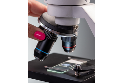

The objectives are one of the most important components within a microscope as they are responsible for image formation and quality.
But what does the objective lens actually do, and should I be thinking of adding oil immersion objectives to my school microscopes, for KS3 4 and 5?
School Microscopes: What do The Numbers on an Objective Mean?

Magnification
Objectives are a key component in magnifying the specimen you are looking at. The magnification power of an objective for a typical educational microscope would be between 4x and 100x. (this will be further magnified by the eyepiece which tends to be 10x).
Navigating Microscope Numerical Aperture - NA
The numerical aperture is related to the angles of light that are collected in a lens which in turn affects the image resolution, the ability to distinguish details within the specimen you are viewing. The higher the NA value the better the resolution.
Please note that the numerical aperture also relates to the condenser. The numerical aperture of the condenser must equal or exceed the numerical aperture of the objective.
For example, the objective pictured above has a numerical aperture of 1.25 and would be best suited to use on a microscope with an Abbe condenser which will offer a numerical aperture of 1.25 too. It would not be advisable to use this objective on a microscope with a 0.65 NA condenser as the best results would not be achieved.
What is the Objective Tube Length?
This is the distance from the opening of the nosepiece, where the objective is screwed in, to the top of the observation tube, where you insert the eyepiece.
If this distance was lengthened, for example by adding an accessory like a polarizer, then the image would be distorted. To use accessories, you need to have objectives that are marked as infinity-corrected.

Choosing the Correct Thickness of Cover Slips
A coverslip is usually made from thin borosilicate glass and is used to cover specimens to be viewed on a microscope slide.
The standard thickness for a cover slip is 0.17 mm, this is a number 1½ cover slip. Higher numbers indicate thicker glass. For optimum results use the thickness of coverslip identified on your microscope objective.
You can view our extensive microscopy range here.
Explaining ‘Achromatic’ Objectives in School Microscopes
This is the most common type of objective.
An achromatic objective limits the effects of chromatic and spherical aberration (the way the rays of light are focused in the focal point).
Light is broken down into various wavelengths as it travels through a lens. These various wavelengths have different focal points, meaning that red, green, and blue appear to focus at different points. This can mean that objects appear to have coloured edges around them and are not as sharp as we might expect (chromatic aberration).

Achromatic objectives correct for colour by bringing the blue and red, into common focus. For higher level microscopy, an apochromatic objective also includes additional correction for green.
Book your FREE microscope demo for expert advice and demonstations.
Improving Flat Field Quality
In addition to colour correction, an achromatic objective offers a flat field of about 65% of the centre of the image. As you work through Semi-Plan, E-Plan and Plan objectives, the percentage of flat field increases.

Oil Immersion Techniques for Enhancing Microscopy
Oil immersion is a technique used in microscopy to increase the resolution and clarity of images. A special oil is used to fill the gap between the microscope objective lens and the sample on the microscope slide. It improves the light-gathering ability of the microscope and allows for sharper images and more accurate measurements.
Oil immersion is not compatible with all microscope objectives and a special ‘oil immersion objective’ is required. These can be easily screwed into the revolving nosepiece of a suitable microscope.
Want to Find out More?
Book your FREE microscope demo from industry experts.